gsl ib4 conjugated to fitc Search Results


95
Vector Laboratories isolectin b4
Isolectin B4, supplied by Vector Laboratories, used in various techniques. Bioz Stars score: 95/100, based on 1 PubMed citations. ZERO BIAS - scores, article reviews, protocol conditions and more
https://www.bioz.com/result/isolectin b4/product/Vector Laboratories
Average 95 stars, based on 1 article reviews
isolectin b4 - by Bioz Stars, 2026-03
95/100 stars
  Buy from Supplier

96
Vector Laboratories isolectin b4 conjugated with biotin
Isolectin B4 Conjugated With Biotin, supplied by Vector Laboratories, used in various techniques. Bioz Stars score: 96/100, based on 1 PubMed citations. ZERO BIAS - scores, article reviews, protocol conditions and more
https://www.bioz.com/result/isolectin b4 conjugated with biotin/product/Vector Laboratories
Average 96 stars, based on 1 article reviews
isolectin b4 conjugated with biotin - by Bioz Stars, 2026-03
96/100 stars
  Buy from Supplier

94
Vector Laboratories isolectin b4 conjugated to fluorescein
Isolectin B4 Conjugated To Fluorescein, supplied by Vector Laboratories, used in various techniques. Bioz Stars score: 94/100, based on 1 PubMed citations. ZERO BIAS - scores, article reviews, protocol conditions and more
https://www.bioz.com/result/isolectin b4 conjugated to fluorescein/product/Vector Laboratories
Average 94 stars, based on 1 article reviews
isolectin b4 conjugated to fluorescein - by Bioz Stars, 2026-03
94/100 stars
  Buy from Supplier

90
Thermo Fisher alexafluor 647 conjugated gsl ib4 lectin
Alexafluor 647 Conjugated Gsl Ib4 Lectin, supplied by Thermo Fisher, used in various techniques. Bioz Stars score: 90/100, based on 1 PubMed citations. ZERO BIAS - scores, article reviews, protocol conditions and more
https://www.bioz.com/result/alexafluor 647 conjugated gsl ib4 lectin/product/Thermo Fisher
Average 90 stars, based on 1 article reviews
alexafluor 647 conjugated gsl ib4 lectin - by Bioz Stars, 2026-03
90/100 stars
  Buy from Supplier

95
Vector Laboratories fitc conjugated ib 4
L4 dorsal root ganglion (DRG) immunostaining for <t>IB-4</t> (a) and CGRP (b) after SNI in male (M) and female (F) rats and representative images of staining in L4 (c). **** p < 0.001, ** p < 0.01 by Student’s t -test with Holm-Sidak correction for multiple comparisons and ANOVA. ## p < 0.01 by ANOVA. n = 10
Fitc Conjugated Ib 4, supplied by Vector Laboratories, used in various techniques. Bioz Stars score: 95/100, based on 1 PubMed citations. ZERO BIAS - scores, article reviews, protocol conditions and more
https://www.bioz.com/result/fitc conjugated ib 4/product/Vector Laboratories
Average 95 stars, based on 1 article reviews
fitc conjugated ib 4 - by Bioz Stars, 2026-03
95/100 stars
  Buy from Supplier

94
Vector Laboratories dylight594 conjugated gsl ib4
L4 dorsal root ganglion (DRG) immunostaining for <t>IB-4</t> (a) and CGRP (b) after SNI in male (M) and female (F) rats and representative images of staining in L4 (c). **** p < 0.001, ** p < 0.01 by Student’s t -test with Holm-Sidak correction for multiple comparisons and ANOVA. ## p < 0.01 by ANOVA. n = 10
Dylight594 Conjugated Gsl Ib4, supplied by Vector Laboratories, used in various techniques. Bioz Stars score: 94/100, based on 1 PubMed citations. ZERO BIAS - scores, article reviews, protocol conditions and more
https://www.bioz.com/result/dylight594 conjugated gsl ib4/product/Vector Laboratories
Average 94 stars, based on 1 article reviews
dylight594 conjugated gsl ib4 - by Bioz Stars, 2026-03
94/100 stars
  Buy from Supplier

95
Vector Laboratories dylight649 conjugated gsl ib4 lectin
L4 dorsal root ganglion (DRG) immunostaining for <t>IB-4</t> (a) and CGRP (b) after SNI in male (M) and female (F) rats and representative images of staining in L4 (c). **** p < 0.001, ** p < 0.01 by Student’s t -test with Holm-Sidak correction for multiple comparisons and ANOVA. ## p < 0.01 by ANOVA. n = 10
Dylight649 Conjugated Gsl Ib4 Lectin, supplied by Vector Laboratories, used in various techniques. Bioz Stars score: 95/100, based on 1 PubMed citations. ZERO BIAS - scores, article reviews, protocol conditions and more
https://www.bioz.com/result/dylight649 conjugated gsl ib4 lectin/product/Vector Laboratories
Average 95 stars, based on 1 article reviews
dylight649 conjugated gsl ib4 lectin - by Bioz Stars, 2026-03
95/100 stars
  Buy from Supplier

95
Vector Laboratories biotin conjugated griffonia
To better define the lineages of infected cells, we stained paraffin sections with cell-specific markers in conjunction with ISH for influenza viral RNA. (A to C) Sections stained with <t>Griffonia</t> simplicifolia lectin that binds to ferret macrophages. (D) Section stained with anti-cytokeratin antibody that binds to epithelial cells. (A and B) Small bowel sections show infected cells (circled) in the lamina propria that display both peroxidase reaction product (red) and ISH grains (black), indicating infection of lamina propria macrophages. (C) Liver section demonstrates infected macrophage elements in regions of periportal hepatitis. (D) Some bile duct epithelial cells labeled for cytokeratin (red) also hybridize with the influenza virus probe (black grains). (E and F) Double-label immunofluorescent images illustrate inflamed liver bile duct epithelia stained for cytokeratin (red) that colocalize with H5 HA protein (green) (E) and infected neurons that stain with microtubule-associated protein-2 (red) and influenza virus H5 HA protein (green) (F).
Biotin Conjugated Griffonia, supplied by Vector Laboratories, used in various techniques. Bioz Stars score: 95/100, based on 1 PubMed citations. ZERO BIAS - scores, article reviews, protocol conditions and more
https://www.bioz.com/result/biotin conjugated griffonia/product/Vector Laboratories
Average 95 stars, based on 1 article reviews
biotin conjugated griffonia - by Bioz Stars, 2026-03
95/100 stars
  Buy from Supplier

95
Vector Laboratories bandeiraea simplicifolia isolectin b4 ib4 conjugated to rhodamine
a , SARS-CoV-2 RNA was quantified by RT-qPCR separately in SCG, TG, and DRG neurons and media to generate a 4–5-day viral genome replication profile in neurons from hACE2 and WT mice. Viral genome replication occurs in all neurons following similar patterns with differential infectious virus release into media. Intracellular replication patterns are similar between hACE2 and WT neurons, although at reduced levels in WT neurons. hACE2 DRGs have peaks in genome replication ~48 hpi and ~96 hpi indicating successive rounds of replication. b , Infectious virus was quantified by plaque assay on Vero E6 cells in SCG, TG, and DRG neuronal cultures to generate growth curves in primary neurons from hACE2 and WT mice. Infectious virus was not recovered from SCG neurons indicating abortive infection, likely mediated by cytotoxicity. Infectious virus was recovered from TG and DRG neurons indicating productive infection of these neurons. c , Immunofluorescence for SARS-N (grey) and either tyrosine hydroxylase (TH) or <t>Isolectin-B4</t> <t>(IB4)</t> to counterstain neurons, or glutamine synthase (GS) to stain satellite glial cells. SARS-N was observed in neurons from each of the ganglia. Infected neurons were largely free of neurites by 1 dpi. At 3 dpi, many infected neurons exhibited cytopathologies such as degraded neurites, enlarged multi-nucleated cell bodies (arrow) compared to uninfected neurons (arrowhead), and SARS-N+ puncta reminiscent of viral replication compartments. See Supplementary Video 3 for 3D rendering of DRG at 3 dpi. See Supplementary Video 4 for 3D rendering of TG at 2 dpi. d, The percentage of hACE2 autonomic (SCG) and sensory (DRG) neurons positive for SARS-N were counted from 1-3 dpi. A small percentage of autonomic (SCG) neurons were visibly infected, with significant observable cell death, similar to in vivo observations. Infection in sensory (DRG) neurons were consistent from 1 −3 dpi, with ~5% infected. Infection of ex vivo neurons is less efficient than in vivo infection. Scale bar = 20 μm. Data are the mean ± s.e.m
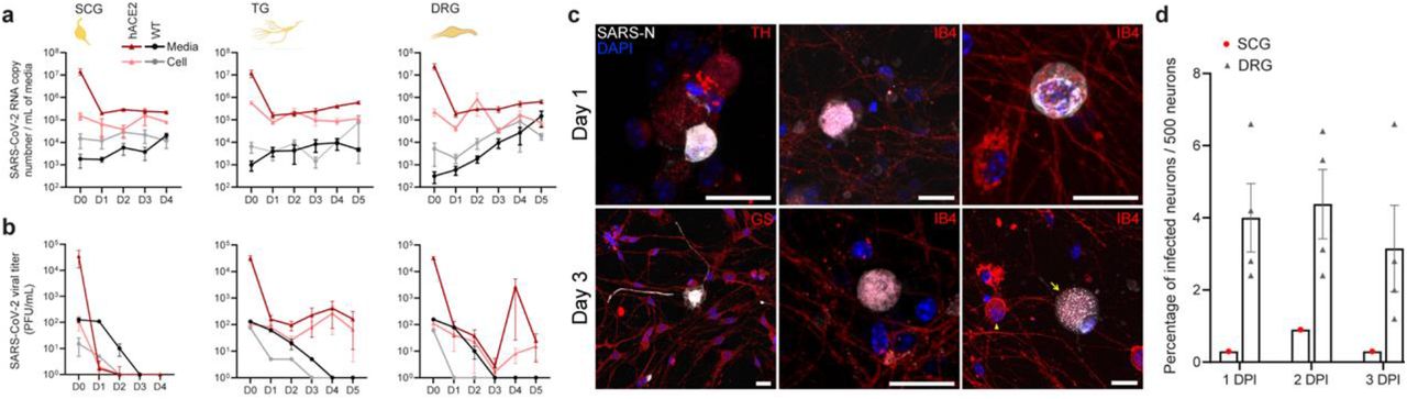
Bandeiraea Simplicifolia Isolectin B4 Ib4 Conjugated To Rhodamine, supplied by Vector Laboratories, used in various techniques. Bioz Stars score: 95/100, based on 1 PubMed citations. ZERO BIAS - scores, article reviews, protocol conditions and more
https://www.bioz.com/result/bandeiraea simplicifolia isolectin b4 ib4 conjugated to rhodamine/product/Vector Laboratories
Average 95 stars, based on 1 article reviews
bandeiraea simplicifolia isolectin b4 ib4 conjugated to rhodamine - by Bioz Stars, 2026-03
95/100 stars
  Buy from Supplier

96
Vector Laboratories biotin conjugated isolectin b4 ib4
a , SARS-CoV-2 RNA was quantified by RT-qPCR separately in SCG, TG, and DRG neurons and media to generate a 4–5-day viral genome replication profile in neurons from hACE2 and WT mice. Viral genome replication occurs in all neurons following similar patterns with differential infectious virus release into media. Intracellular replication patterns are similar between hACE2 and WT neurons, although at reduced levels in WT neurons. hACE2 DRGs have peaks in genome replication ~48 hpi and ~96 hpi indicating successive rounds of replication. b , Infectious virus was quantified by plaque assay on Vero E6 cells in SCG, TG, and DRG neuronal cultures to generate growth curves in primary neurons from hACE2 and WT mice. Infectious virus was not recovered from SCG neurons indicating abortive infection, likely mediated by cytotoxicity. Infectious virus was recovered from TG and DRG neurons indicating productive infection of these neurons. c , Immunofluorescence for SARS-N (grey) and either tyrosine hydroxylase (TH) or <t>Isolectin-B4</t> <t>(IB4)</t> to counterstain neurons, or glutamine synthase (GS) to stain satellite glial cells. SARS-N was observed in neurons from each of the ganglia. Infected neurons were largely free of neurites by 1 dpi. At 3 dpi, many infected neurons exhibited cytopathologies such as degraded neurites, enlarged multi-nucleated cell bodies (arrow) compared to uninfected neurons (arrowhead), and SARS-N+ puncta reminiscent of viral replication compartments. See Supplementary Video 3 for 3D rendering of DRG at 3 dpi. See Supplementary Video 4 for 3D rendering of TG at 2 dpi. d, The percentage of hACE2 autonomic (SCG) and sensory (DRG) neurons positive for SARS-N were counted from 1-3 dpi. A small percentage of autonomic (SCG) neurons were visibly infected, with significant observable cell death, similar to in vivo observations. Infection in sensory (DRG) neurons were consistent from 1 −3 dpi, with ~5% infected. Infection of ex vivo neurons is less efficient than in vivo infection. Scale bar = 20 μm. Data are the mean ± s.e.m
Biotin Conjugated Isolectin B4 Ib4, supplied by Vector Laboratories, used in various techniques. Bioz Stars score: 96/100, based on 1 PubMed citations. ZERO BIAS - scores, article reviews, protocol conditions and more
https://www.bioz.com/result/biotin conjugated isolectin b4 ib4/product/Vector Laboratories
Average 96 stars, based on 1 article reviews
biotin conjugated isolectin b4 ib4 - by Bioz Stars, 2026-03
96/100 stars
  Buy from Supplier

92
Vector Laboratories griffonia simplicifolia
a , SARS-CoV-2 RNA was quantified by RT-qPCR separately in SCG, TG, and DRG neurons and media to generate a 4–5-day viral genome replication profile in neurons from hACE2 and WT mice. Viral genome replication occurs in all neurons following similar patterns with differential infectious virus release into media. Intracellular replication patterns are similar between hACE2 and WT neurons, although at reduced levels in WT neurons. hACE2 DRGs have peaks in genome replication ~48 hpi and ~96 hpi indicating successive rounds of replication. b , Infectious virus was quantified by plaque assay on Vero E6 cells in SCG, TG, and DRG neuronal cultures to generate growth curves in primary neurons from hACE2 and WT mice. Infectious virus was not recovered from SCG neurons indicating abortive infection, likely mediated by cytotoxicity. Infectious virus was recovered from TG and DRG neurons indicating productive infection of these neurons. c , Immunofluorescence for SARS-N (grey) and either tyrosine hydroxylase (TH) or <t>Isolectin-B4</t> <t>(IB4)</t> to counterstain neurons, or glutamine synthase (GS) to stain satellite glial cells. SARS-N was observed in neurons from each of the ganglia. Infected neurons were largely free of neurites by 1 dpi. At 3 dpi, many infected neurons exhibited cytopathologies such as degraded neurites, enlarged multi-nucleated cell bodies (arrow) compared to uninfected neurons (arrowhead), and SARS-N+ puncta reminiscent of viral replication compartments. See Supplementary Video 3 for 3D rendering of DRG at 3 dpi. See Supplementary Video 4 for 3D rendering of TG at 2 dpi. d, The percentage of hACE2 autonomic (SCG) and sensory (DRG) neurons positive for SARS-N were counted from 1-3 dpi. A small percentage of autonomic (SCG) neurons were visibly infected, with significant observable cell death, similar to in vivo observations. Infection in sensory (DRG) neurons were consistent from 1 −3 dpi, with ~5% infected. Infection of ex vivo neurons is less efficient than in vivo infection. Scale bar = 20 μm. Data are the mean ± s.e.m
Griffonia Simplicifolia, supplied by Vector Laboratories, used in various techniques. Bioz Stars score: 92/100, based on 1 PubMed citations. ZERO BIAS - scores, article reviews, protocol conditions and more
https://www.bioz.com/result/griffonia simplicifolia/product/Vector Laboratories
Average 92 stars, based on 1 article reviews
griffonia simplicifolia - by Bioz Stars, 2026-03
92/100 stars
  Buy from Supplier

Image Search Results


L4 dorsal root ganglion (DRG) immunostaining for IB-4 (a) and CGRP (b) after SNI in male (M) and female (F) rats and representative images of staining in L4 (c). **** p < 0.001, ** p < 0.01 by Student’s t -test with Holm-Sidak correction for multiple comparisons and ANOVA. ## p < 0.01 by ANOVA. n = 10

Journal: Molecular Neurobiology

Article Title: Spared Nerve Injury Causes Sexually Dimorphic Mechanical Allodynia and Differential Gene Expression in Spinal Cords and Dorsal Root Ganglia in Rats

doi: 10.1007/s12035-021-02447-1

Figure Lengend Snippet: L4 dorsal root ganglion (DRG) immunostaining for IB-4 (a) and CGRP (b) after SNI in male (M) and female (F) rats and representative images of staining in L4 (c). **** p < 0.001, ** p < 0.01 by Student’s t -test with Holm-Sidak correction for multiple comparisons and ANOVA. ## p < 0.01 by ANOVA. n = 10

Article Snippet: DRG sections were probed with antibodies for CGRP (1:10,000, Cat# T-4032, Peninsula Laboratories, San Carlos, CA, USA) or FITC-conjugated IB-4 (1:200; Cat# FL-1201, Vector Labs, CA, USA).

Techniques: Immunostaining, Staining

To better define the lineages of infected cells, we stained paraffin sections with cell-specific markers in conjunction with ISH for influenza viral RNA. (A to C) Sections stained with Griffonia simplicifolia lectin that binds to ferret macrophages. (D) Section stained with anti-cytokeratin antibody that binds to epithelial cells. (A and B) Small bowel sections show infected cells (circled) in the lamina propria that display both peroxidase reaction product (red) and ISH grains (black), indicating infection of lamina propria macrophages. (C) Liver section demonstrates infected macrophage elements in regions of periportal hepatitis. (D) Some bile duct epithelial cells labeled for cytokeratin (red) also hybridize with the influenza virus probe (black grains). (E and F) Double-label immunofluorescent images illustrate inflamed liver bile duct epithelia stained for cytokeratin (red) that colocalize with H5 HA protein (green) (E) and infected neurons that stain with microtubule-associated protein-2 (red) and influenza virus H5 HA protein (green) (F).

Journal: Journal of Virology

Article Title: H1N1, but Not H3N2, Influenza A Virus Infection Protects Ferrets from H5N1 Encephalitis

doi: 10.1128/JVI.01840-13

Figure Lengend Snippet: To better define the lineages of infected cells, we stained paraffin sections with cell-specific markers in conjunction with ISH for influenza viral RNA. (A to C) Sections stained with Griffonia simplicifolia lectin that binds to ferret macrophages. (D) Section stained with anti-cytokeratin antibody that binds to epithelial cells. (A and B) Small bowel sections show infected cells (circled) in the lamina propria that display both peroxidase reaction product (red) and ISH grains (black), indicating infection of lamina propria macrophages. (C) Liver section demonstrates infected macrophage elements in regions of periportal hepatitis. (D) Some bile duct epithelial cells labeled for cytokeratin (red) also hybridize with the influenza virus probe (black grains). (E and F) Double-label immunofluorescent images illustrate inflamed liver bile duct epithelia stained for cytokeratin (red) that colocalize with H5 HA protein (green) (E) and infected neurons that stain with microtubule-associated protein-2 (red) and influenza virus H5 HA protein (green) (F).

Article Snippet: For lectin histochemistry to detect ferret macrophages ( 38 ), sections were incubated with biotin-conjugated Griffonia ( Bandeiraea ) simplicifolia isolectin B 4 (Vector Laboratories, Burlingame, CA) instead of antibodies.

Techniques: Infection, Staining, Labeling

a , SARS-CoV-2 RNA was quantified by RT-qPCR separately in SCG, TG, and DRG neurons and media to generate a 4–5-day viral genome replication profile in neurons from hACE2 and WT mice. Viral genome replication occurs in all neurons following similar patterns with differential infectious virus release into media. Intracellular replication patterns are similar between hACE2 and WT neurons, although at reduced levels in WT neurons. hACE2 DRGs have peaks in genome replication ~48 hpi and ~96 hpi indicating successive rounds of replication. b , Infectious virus was quantified by plaque assay on Vero E6 cells in SCG, TG, and DRG neuronal cultures to generate growth curves in primary neurons from hACE2 and WT mice. Infectious virus was not recovered from SCG neurons indicating abortive infection, likely mediated by cytotoxicity. Infectious virus was recovered from TG and DRG neurons indicating productive infection of these neurons. c , Immunofluorescence for SARS-N (grey) and either tyrosine hydroxylase (TH) or Isolectin-B4 (IB4) to counterstain neurons, or glutamine synthase (GS) to stain satellite glial cells. SARS-N was observed in neurons from each of the ganglia. Infected neurons were largely free of neurites by 1 dpi. At 3 dpi, many infected neurons exhibited cytopathologies such as degraded neurites, enlarged multi-nucleated cell bodies (arrow) compared to uninfected neurons (arrowhead), and SARS-N+ puncta reminiscent of viral replication compartments. See Supplementary Video 3 for 3D rendering of DRG at 3 dpi. See Supplementary Video 4 for 3D rendering of TG at 2 dpi. d, The percentage of hACE2 autonomic (SCG) and sensory (DRG) neurons positive for SARS-N were counted from 1-3 dpi. A small percentage of autonomic (SCG) neurons were visibly infected, with significant observable cell death, similar to in vivo observations. Infection in sensory (DRG) neurons were consistent from 1 −3 dpi, with ~5% infected. Infection of ex vivo neurons is less efficient than in vivo infection. Scale bar = 20 μm. Data are the mean ± s.e.m

Journal: bioRxiv

Article Title: SARS-CoV-2 Infects Peripheral and Central Neurons of Mice Before Viremia, Facilitated by Neuropilin-1

doi: 10.1101/2022.05.20.492834

Figure Lengend Snippet: a , SARS-CoV-2 RNA was quantified by RT-qPCR separately in SCG, TG, and DRG neurons and media to generate a 4–5-day viral genome replication profile in neurons from hACE2 and WT mice. Viral genome replication occurs in all neurons following similar patterns with differential infectious virus release into media. Intracellular replication patterns are similar between hACE2 and WT neurons, although at reduced levels in WT neurons. hACE2 DRGs have peaks in genome replication ~48 hpi and ~96 hpi indicating successive rounds of replication. b , Infectious virus was quantified by plaque assay on Vero E6 cells in SCG, TG, and DRG neuronal cultures to generate growth curves in primary neurons from hACE2 and WT mice. Infectious virus was not recovered from SCG neurons indicating abortive infection, likely mediated by cytotoxicity. Infectious virus was recovered from TG and DRG neurons indicating productive infection of these neurons. c , Immunofluorescence for SARS-N (grey) and either tyrosine hydroxylase (TH) or Isolectin-B4 (IB4) to counterstain neurons, or glutamine synthase (GS) to stain satellite glial cells. SARS-N was observed in neurons from each of the ganglia. Infected neurons were largely free of neurites by 1 dpi. At 3 dpi, many infected neurons exhibited cytopathologies such as degraded neurites, enlarged multi-nucleated cell bodies (arrow) compared to uninfected neurons (arrowhead), and SARS-N+ puncta reminiscent of viral replication compartments. See Supplementary Video 3 for 3D rendering of DRG at 3 dpi. See Supplementary Video 4 for 3D rendering of TG at 2 dpi. d, The percentage of hACE2 autonomic (SCG) and sensory (DRG) neurons positive for SARS-N were counted from 1-3 dpi. A small percentage of autonomic (SCG) neurons were visibly infected, with significant observable cell death, similar to in vivo observations. Infection in sensory (DRG) neurons were consistent from 1 −3 dpi, with ~5% infected. Infection of ex vivo neurons is less efficient than in vivo infection. Scale bar = 20 μm. Data are the mean ± s.e.m

Article Snippet: NeuN was visualized using an Alexa Fluor ® 647 conjugated rabbit monoclonal anti-NeuN antibody at a 1:1000 concentration (ab190565; Abcam). α-d-galactose carbohydrate residues on sensory neurons was visualized using the Bandeiraea simplicifolia isolectin B4 (IB4) conjugated to rhodamine at a 1:250 concentration (RL-1102; Vector Laboratories).

Techniques: Quantitative RT-PCR, Plaque Assay, Infection, Immunofluorescence, Staining, In Vivo, Ex Vivo

Cells were fixed 1-3 dpi and stained for SARS-N and various counterstains. All images were acquired using a Leica SP8 confocal microscope. Because of substantial variability in intensity of SARS-N immunofluorescence, laser power and gain were adjusted in order to highlight features of each cell. Day 2 DRG is a montage image to show SARS-N detected throughout the neurites of one infected neuron; only DAPI is shown. SARS-N is present in neurons in the SCG, TG, and DRG in infected mice. TH = tyrosine hydroxylase, IB4 = Isolectin-IB4, GS = glutamine synthetase, SARS-N =SARS-CoV-2 nucleocapsid, DAPI = 4’,6-diamidino-2-phenylindole. Scale bar = 20 μm

Journal: bioRxiv

Article Title: SARS-CoV-2 Infects Peripheral and Central Neurons of Mice Before Viremia, Facilitated by Neuropilin-1

doi: 10.1101/2022.05.20.492834

Figure Lengend Snippet: Cells were fixed 1-3 dpi and stained for SARS-N and various counterstains. All images were acquired using a Leica SP8 confocal microscope. Because of substantial variability in intensity of SARS-N immunofluorescence, laser power and gain were adjusted in order to highlight features of each cell. Day 2 DRG is a montage image to show SARS-N detected throughout the neurites of one infected neuron; only DAPI is shown. SARS-N is present in neurons in the SCG, TG, and DRG in infected mice. TH = tyrosine hydroxylase, IB4 = Isolectin-IB4, GS = glutamine synthetase, SARS-N =SARS-CoV-2 nucleocapsid, DAPI = 4’,6-diamidino-2-phenylindole. Scale bar = 20 μm

Article Snippet: NeuN was visualized using an Alexa Fluor ® 647 conjugated rabbit monoclonal anti-NeuN antibody at a 1:1000 concentration (ab190565; Abcam). α-d-galactose carbohydrate residues on sensory neurons was visualized using the Bandeiraea simplicifolia isolectin B4 (IB4) conjugated to rhodamine at a 1:250 concentration (RL-1102; Vector Laboratories).

Techniques: Staining, Microscopy, Immunofluorescence, Infection

Immunofluorescence for SARS-N (grey) and Isolectin-B4 (IB4) to counterstain neurons shows a variety of phenotypes of infected cells, including neurons with a loss of membrane integrity (1), SARS-N+ puncta within and surrounding neurons (2), and seemingly healthy neurons with extensive neurites with strong SARS-N+ staining (arrow in 3). Infected satellite glial cells were also observed (arrowheads in 3); many appeared to be activated, noted by the presence of extended cellular processes. These findings are similar to immunostaining of DRGs in vivo, which also contained numerous infected satellite glial cells.

Journal: bioRxiv

Article Title: SARS-CoV-2 Infects Peripheral and Central Neurons of Mice Before Viremia, Facilitated by Neuropilin-1

doi: 10.1101/2022.05.20.492834

Figure Lengend Snippet: Immunofluorescence for SARS-N (grey) and Isolectin-B4 (IB4) to counterstain neurons shows a variety of phenotypes of infected cells, including neurons with a loss of membrane integrity (1), SARS-N+ puncta within and surrounding neurons (2), and seemingly healthy neurons with extensive neurites with strong SARS-N+ staining (arrow in 3). Infected satellite glial cells were also observed (arrowheads in 3); many appeared to be activated, noted by the presence of extended cellular processes. These findings are similar to immunostaining of DRGs in vivo, which also contained numerous infected satellite glial cells.

Article Snippet: NeuN was visualized using an Alexa Fluor ® 647 conjugated rabbit monoclonal anti-NeuN antibody at a 1:1000 concentration (ab190565; Abcam). α-d-galactose carbohydrate residues on sensory neurons was visualized using the Bandeiraea simplicifolia isolectin B4 (IB4) conjugated to rhodamine at a 1:250 concentration (RL-1102; Vector Laboratories).

Techniques: Immunofluorescence, Infection, Staining, Immunostaining, In Vivo